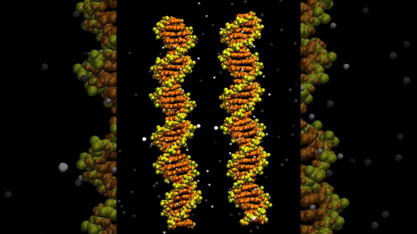
DNA needs a little help to zip up its genes.
In a first, scientists have visualized how pieces of DNA pair up. Computer simulations further reveal that charged metal ions help twin double helices align, researchers report September 9 in Nucleic Acids Research.
The work adds insight to the process in which cells pair up chromosomes before divvying DNA into eggs or sperm. Matching the chromosomes allows them to exchange information and keeps them from going astray, much like how folding socks keeps pairs together.
But it is not so easy to bring two DNA double helices together. The strands have a negative electrical charge and repel each other. Researchers previously hypothesized that positively charged ions could facilitate zipping the twisting strands of DNA together.
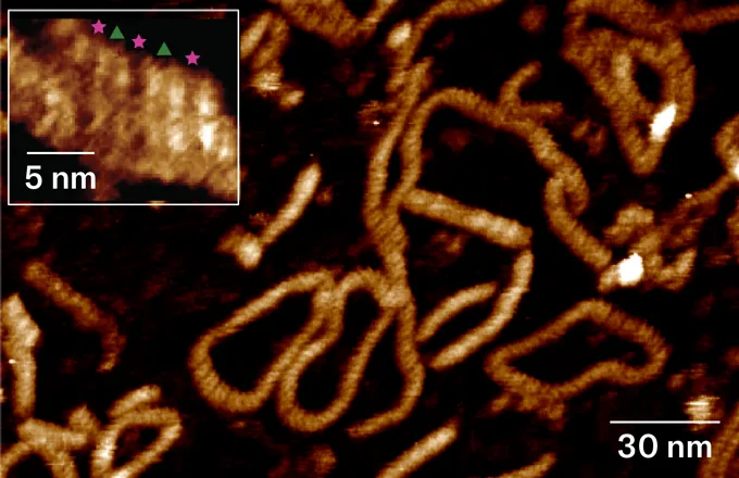
Scientists at the University of Sheffield in England used atomic force microscopy to visualize short strands of DNA pairing up. The specialized microscope technique works like a record player, says microscopist Thomas Catley. A very sharp-tipped needle is dragged across a surface containing molecules the researchers want to examine. "We detect how that tip moves, and we turn that into an image signal, basically the same way that you get sound from a record player," he says.
The zipped DNA strands look like twisted pieces of yarn with their grooves nestling together.
DNA pairing revealed
An atomic force microscope image shows identical DNA double helices pairing up. A zoomed in look (box, upper left) shows two double strands of DNA nestling together. Pink stars and green triangles indicate the alternating arrangement of grooves where the attraction between positively charged metal ions and negatively charged DNA holds the helices together.
Agnes Noy, a computational biophysicist at the University of York in England, was so taken with the images that she and colleagues did computer simulations to explain the mechanism. Positively charged metal ions nestle in the grooves of the DNA's spiral staircases. That's where information-carrying bases sit. The ions form bridges with the negatively charged ridges of the DNA backbone.
The DNA strands are staggered so that positive and negative charges alternate, the simulations suggest. The opposite charges attract the strands to each other. Together the images and simulations confirm the DNA zipper model, the researchers say.
This knowledge may help bioengineers make better folds in DNA origami structures used to deliver drugs and give a better understanding of biological processes, including what goes wrong in some cancers.

Comments 0
No comments yet. Start the conversation.
Leave a comment